


When Is Ultrasound Done in Pregnancy: A Complete Trimester-Wise Guide

Summary
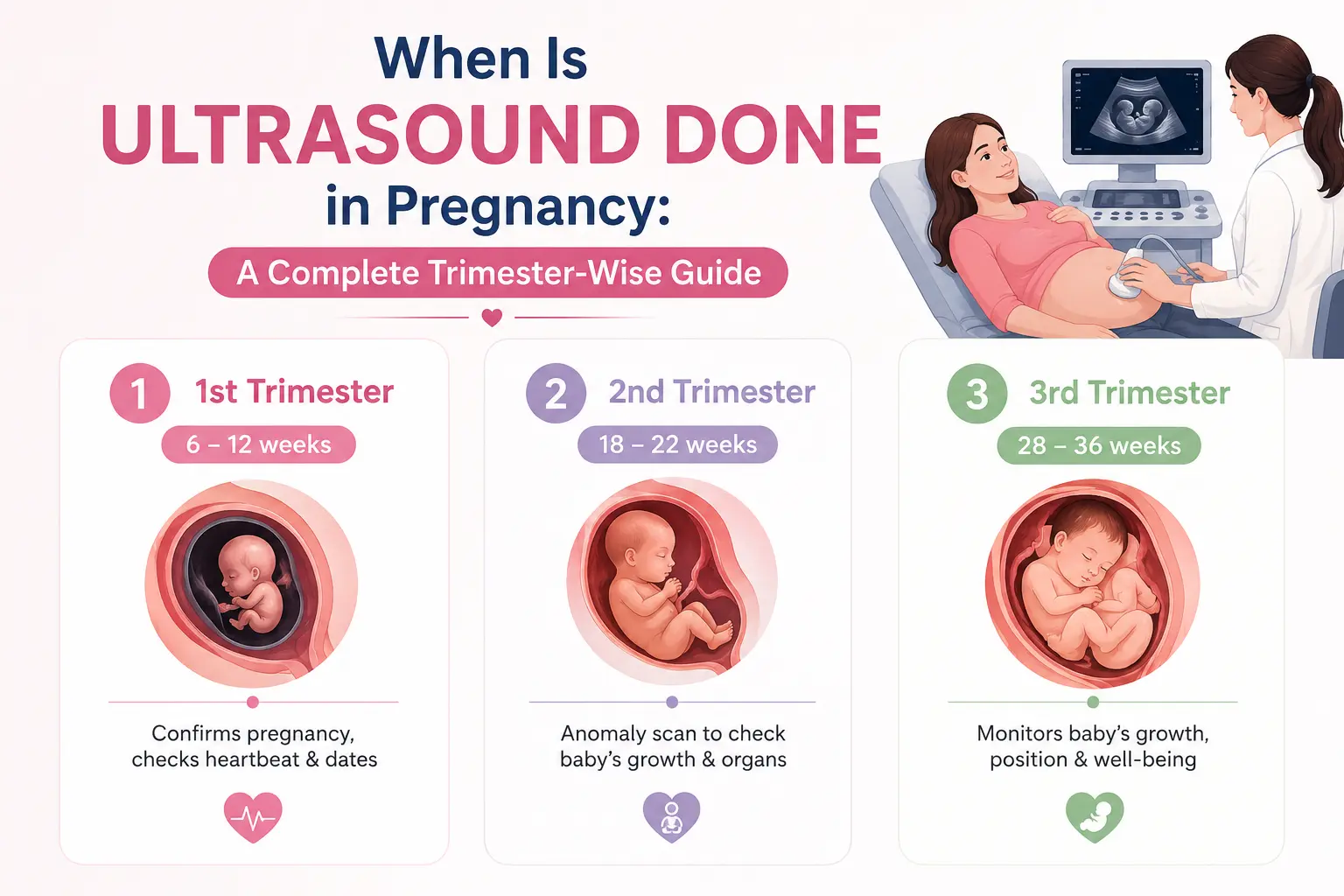
Pregnancy is a journey full of firsts - and one of the most reassuring milestones for every expectant mother is seeing her baby for the first time on an ultrasound screen. But beyond that emotional moment, pregnancy ultrasounds serve a vital clinical purpose at every stage. Knowing when an ultrasound is done in pregnancy, what each scan looks for, and what results mean can help mothers approach every appointment with clarity and confidence.
This complete guide walks you through the full pregnancy ultrasound schedule - trimester by trimester - covering the purpose of each scan, what doctors look for, and when additional scans may be necessary.
What Is a Pregnancy Ultrasound?
A pregnancy ultrasound, also called a sonography or USG scan, uses high-frequency sound waves to produce real-time images of the baby, placenta, amniotic fluid, and uterus. It is completely safe, painless, and uses no radiation - making it the preferred imaging method throughout pregnancy.
Ultrasound in pregnancy serves multiple purposes at different stages:
-
Confirming pregnancy and estimating gestational age
-
Detecting the foetal heartbeat
-
Screening for chromosomal abnormalities
-
Assessing foetal growth, position, and wellbeing
-
Evaluating the placenta and amniotic fluid levels
-
Detecting structural abnormalities in the developing baby
-
Guiding clinical decisions for high-risk pregnancies
At Miracles Healthcare, ultrasound scans are performed by experienced, NABL-certified radiologists - including dedicated female radiologists - with reports delivered the same day, directly to your phone via WhatsApp.
First Trimester Ultrasounds (Weeks 1-13)
Early Pregnancy Scan - Weeks 6 to 8
The first ultrasound in pregnancy is typically done between 6 and 8 weeks - sometimes as early as week 5 if there are specific concerns. At this early stage, the uterus is still small, which is why this scan is usually performed as a TVS (Transvaginal Ultrasound), which uses a small probe inserted into the vagina for closer and clearer imaging of the uterus and ovaries.
What this scan checks:
-
Confirms an intrauterine pregnancy (rules out ectopic pregnancy)
-
Detects the foetal heartbeat, typically visible from week 6 onwards
-
Establishes gestational age and estimates the due date
-
Identifies the number of embryos (singleton, twins, or multiples)
-
Evaluates the uterus and ovaries for any abnormalities, such as fibroids or cysts
It is important to understand that if a heartbeat is not detected before 7 weeks, this does not necessarily indicate a problem - the embryo may simply be too small to detect clearly. A follow-up scan is usually advised within 1-2 weeks.
Nuchal Translucency (NT) Scan - Weeks 11 to 14
The NT scan is one of the most significant scans of the first trimester. It is done between 11 weeks and 13 weeks 6 days, and measures the fluid at the back of the baby's neck - called nuchal translucency.
What this scan checks:
-
Screening for chromosomal conditions, including Down syndrome (Trisomy 21), Edwards syndrome (Trisomy 18), and Patau syndrome (Trisomy 13)
-
Early assessment of foetal anatomy - heart, brain, spine, and limbs
-
Confirmation of gestational age and due date
-
Foetal nasal bone assessment (absence may indicate chromosomal risk)
The NT scan is usually combined with a blood test (the Double Marker Test) to provide a combined first-trimester risk assessment. This combination significantly improves the accuracy of chromosomal screening.
Second Trimester Ultrasounds (Weeks 14-27)
Level 2 Anomaly Scan - Weeks 18 to 20
The Level 2 Ultrasound - also called the anomaly scan or mid-pregnancy scan - is the most detailed and comprehensive ultrasound done during pregnancy. It is performed between 18 and 20 weeks, when the baby is large enough to be examined in detail but still small enough to be seen in full on screen.
What this scan checks:
-
Detailed structural assessment of the baby's brain, heart, spine, kidneys, abdomen, limbs, and face
-
Detection of foetal abnormalities such as neural tube defects, cleft lip, heart defects, and limb abnormalities
-
Placental position - identifying placenta previa (low-lying placenta) that may require monitoring
-
Amniotic fluid levels - too much (polyhydramnios) or too little (oligohydramnios) can signal problems
-
Foetal growth and measurements, including head circumference, abdominal circumference, and femur length
-
Cervical length - a short cervix may indicate risk of preterm labour
This scan is strongly recommended for all pregnant women. For those with high-risk pregnancies, it is an essential part of the monitoring protocol.
Growth Scan - Weeks 24 to 28 (if required)
Some doctors recommend an additional growth scan in the mid-second trimester, particularly for women with gestational diabetes, hypertension, or a history of small-for-gestational-age babies. This scan monitors foetal growth velocity and checks amniotic fluid levels.
Third Trimester Ultrasounds (Weeks 28-40)
Growth and Wellbeing Scan - Weeks 28 to 32
The third-trimester ultrasound is focused primarily on foetal growth, placental function, and well-being. Most women have at least one scan in this trimester, usually between weeks 28 and 32.
What this scan checks:
-
Foetal growth measurements and estimated foetal weight
-
Amniotic fluid index (AFI) - assessing whether fluid levels are within the normal range
-
Placental position and grading - checking whether a previously identified low-lying placenta has moved
-
Foetal presentation - whether the baby is head-down, breech, or transverse
-
Umbilical cord blood flow via Doppler - assessing oxygen and nutrient delivery to the baby
A Colour Doppler Ultrasound may be added to this scan for women with pregnancy complications, as it provides detailed information about blood flow in the umbilical cord, uterine arteries, and foetal vessels.
Pre-Delivery Scan - Weeks 36 to 40
The final ultrasound before delivery is done in the last few weeks of pregnancy to confirm everything is in order before labour begins.
What this scan checks:
-
Final confirmation of foetal position (head-down is ideal for normal delivery)
-
Estimation of foetal weight to guide delivery planning
-
Placental grade and position - ensuring it is not blocking the cervical opening
-
Amniotic fluid levels - low fluid at term may indicate the need for induction
-
Foetal movements and well-being assessment
For women planning a normal delivery, this scan helps confirm that the baby is in the optimal position. If the baby is in a breech position, a caesarean delivery may be recommended.
How Many Ultrasounds Are Done in a Normal Pregnancy?
In a low-risk, uncomplicated pregnancy, a minimum of three to four ultrasounds is typically recommended:
|
Scan |
Timing |
Purpose |
|
Early Pregnancy / TVS Scan |
6-8 weeks |
Confirm pregnancy, heartbeat, and due date |
|
NT Scan |
11-14 weeks |
Chromosomal screening, foetal anatomy check |
|
Level 2 Anomaly Scan |
18-20 weeks |
Detailed structural assessment |
|
Growth and Wellbeing Scan |
28-32 weeks |
Foetal growth, placenta, fluid levels |
|
Pre-Delivery Scan |
36-40 weeks |
Foetal position, weight, and delivery planning |
Women with high-risk pregnancies - including those with gestational diabetes, hypertension, twins, or a history of pregnancy loss - may require additional ultrasounds every 2 to 4 weeks from the second trimester onwards.
When Are Additional Ultrasounds Needed?
Beyond the routine schedule, your doctor may recommend extra ultrasounds if:
-
You experience bleeding or spotting at any stage of pregnancy
-
Foetal movements reduce significantly after 28 weeks
-
A previous scan raised concerns about foetal growth or anatomy
-
You have a high-risk pregnancy due to a medical condition
-
You are carrying twins or higher-order multiples
-
Your amniotic fluid levels were abnormal on a previous scan
-
You are past your due date, and labour has not yet started (post-term monitoring)
Ultrasound Scans Available at Miracles Healthcare, Gurgaon
Miracles Healthcare - a trusted multi-speciality hospital in Gurgaon serving patients since 2002 - provides the full range of pregnancy ultrasound services through its NABL-certified radiology department. All scans are conducted by experienced radiologists using advanced imaging equipment from leading manufacturers such as GE and Philips. Reports are delivered within 6 hours and shared directly via WhatsApp.
The following pregnancy-related ultrasound scans are available across Miracles Healthcare locations:
-
TVS Ultrasound (Transvaginal Scan) - For early pregnancy confirmation, ectopic pregnancy screening, and pelvic assessment
-
Level 2 Ultrasound (Anomaly Scan) - Detailed foetal structural screening at 18-20 weeks
-
Ultrasound (USG) - including obstetric ultrasound, Colour Doppler, and foetal growth scans
-
Radiology Services - Full-spectrum imaging with qualified female radiologists available for patient comfort
Scans are available at:
-
Miracles Mediclinic, Sector 14 - 0124-4797600
-
Miracles Mediclinic, Sector 56 - 906-906-8800 (MRI also available)
-
Miracles Apollo Cradle/Spectra, Sector 82 - 0124-352-8282
-
Miracles Apollo Cradle, Sector 14 - 0124-4797600
All obstetric scans are supported by the Department of Obstetrics and Gynaecology, ensuring that scan findings are reviewed in the full context of each patient's pregnancy care.
Book Your Pregnancy Ultrasound Today
Whether you need your first early pregnancy scan or a detailed Level 2 anomaly scan, Miracles Healthcare offers expert imaging services with fast reports, experienced radiologists, and easy access across Gurgaon.
Call: 0124-4797600
WhatsApp: 98209-65000
Book online: www.miracleshealth.com
Written and Verified by:

Frequently Asked Questions
The first ultrasound in pregnancy is typically done between 6 and 8 weeks to confirm the pregnancy, detect the foetal heartbeat, and estimate the due date. If a heartbeat is not visible before 7 weeks, a follow-up scan is usually advised within 1 to 2 weeks. For women who have had previous miscarriages or fertility treatments, an early TVS ultrasound may be recommended as early as week 5 or 6.
In a low-risk pregnancy, three to five ultrasounds are typically recommended - at 6-8 weeks, 11-14 weeks, 18-20 weeks, 28-32 weeks, and 36-40 weeks. Women with high-risk conditions such as gestational diabetes, hypertension, or a history of preterm labour may require additional scans every 2 to 4 weeks. Your obstetrics and gynaecology specialist will guide the appropriate scanning schedule for your specific situation.
The Level 2 ultrasound - done at 18 to 20 weeks - is the most comprehensive structural scan of the entire pregnancy. It assesses the baby's brain, heart, spine, kidneys, and limbs in detail, and checks the placenta and amniotic fluid levels. It is the primary tool for detecting foetal abnormalities and should not be skipped, even in low-risk pregnancies.
Yes, pregnancy ultrasound is completely safe for both mother and baby. It uses high-frequency sound waves - not radiation - to produce images, making it the preferred and internationally recommended imaging method throughout all trimesters. There are no known risks associated with routine prenatal ultrasound when performed by qualified radiologists.
A TVS ultrasound (Transvaginal Scan) uses a small probe inserted into the vagina to produce clearer images of the uterus and ovaries during early pregnancy. It is used in the first trimester - typically between 6 and 10 weeks - when the uterus is too small for a clear abdominal scan. It is also used to assess cervical length and detect conditions like placenta previa later in pregnancy.
If a scan identifies a concern - such as a structural abnormality, low amniotic fluid, or abnormal placental position - your doctor will discuss the finding, recommend further investigations if necessary, and guide the appropriate next steps. Many findings require only closer monitoring, while others may prompt referral to a foetal medicine specialist. The fetal medicine team at Miracles Healthcare works alongside the radiology and obstetrics teams for integrated assessment and management.
Yes. Miracles Healthcare provides the full range of pregnancy ultrasound scans - including TVS, Level 2 anomaly scans, Colour Doppler, and growth scans - across its NABL-certified diagnostic centres in Sector 14, Sector 56, and Sector 82. Reports are delivered within 6 hours with WhatsApp delivery, and Sunday scans are available with same-day reporting.







Was the information useful?
0 0